Retinal Detachment: Emergency Symptoms and Surgical Treatment You Can't Afford to Ignore

- Colin Hurd
- 25 December 2025
- 8 Comments
One moment you’re reading a book, the next you see a dark curtain sliding across your vision. Or maybe it’s a sudden storm of floaters-dozens of new specks floating in your eye-that didn’t exist yesterday. You might think it’s just eye strain, fatigue, or aging. But if you’re experiencing these symptoms, you could be facing a retinal detachment-a sight-threatening emergency that demands immediate action.
Retinal detachment happens when the thin, light-sensitive layer at the back of your eye-the retina-pulls away from the tissue that supplies it with oxygen and nutrients. Without prompt treatment, the photoreceptor cells in the retina begin to die. Within hours, vision loss can become permanent. The good news? If you catch it early, surgery can often restore most or all of your vision. The bad news? Most people delay seeking help, and every hour counts.
What Are the Warning Signs?
There are six clear red flags that signal a possible retinal detachment. Recognizing them could save your sight.
- Sudden increase in floaters: Not just one or two. Dozens. Dark spots, squiggly lines, or cobwebs that appear out of nowhere. The National Eye Institute says patients describe this as "a lot of new floaters"-not the occasional one you’ve lived with for years.
- Flashes of light: Bright streaks or sparks, especially in your peripheral vision. These aren’t like the afterimage from a camera flash. They’re sudden, brief, and often happen in the dark or when you move your head.
- A dark curtain or shadow: This is the most urgent sign. It starts in your peripheral vision and creeps inward like a curtain being drawn. Once it reaches the center, your central vision is at risk.
- Blurry or distorted vision: Things look warped, wavy, or out of focus-especially if the macula (the center of the retina) is involved. Studies show this happens in nearly 70% of cases.
- Loss of peripheral vision: You start bumping into things on your left or right. You’re not just "not paying attention." Your side vision is literally disappearing.
- Sudden color changes: Colors look washed out or dull. This usually means the macula is affected. The American Academy of Ophthalmology lists this as a key indicator in their 2023 guidelines.
If you notice even one of these, don’t wait. Don’t call your optometrist tomorrow. Don’t Google it. Go to an emergency eye clinic today. A 2022 study in the Journal of VitreoRetinal Diseases found that patients treated within 24 hours had a 90% chance of successful reattachment. After 72 hours, your chance of regaining 20/40 vision drops from 75% to 35%.
How Is It Diagnosed?
There’s no home test for retinal detachment. You need a specialist with the right tools.
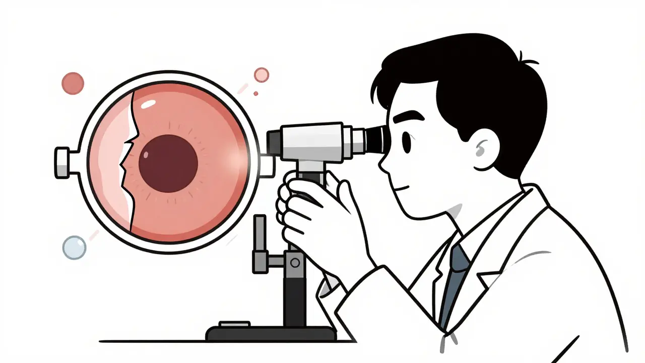
The gold standard is a dilated fundus exam. An ophthalmologist uses a bright light and a special lens (usually a 20D or 90D lens) to look deep into your eye. They can see tears, holes, or areas where the retina has lifted away. If your eye is cloudy from cataracts or bleeding, they’ll use B-scan ultrasound-a painless scan that creates an image of the retina’s position behind the cloudiness.
For detailed imaging, optical coherence tomography (OCT) is used. It’s like a high-res MRI of the retina. It shows exactly how much fluid is under the retina and whether the macula is still attached. This helps surgeons decide if they need to act immediately or if there’s a little more time.
General eye doctors miss about 22% of early detachments, according to a 2022 study. Retinal specialists, who focus only on these conditions, miss only 5%. That’s why if your regular eye doctor says "it’s probably nothing," but you still feel something’s wrong-get a second opinion from a vitreoretinal specialist.
What Are the Surgical Options?
There are three main surgeries. The right one depends on the size, location, and complexity of the detachment.
1. Pneumatic Retinopexy
This is the least invasive option. A gas bubble is injected into the vitreous cavity of your eye. You’re then positioned so the bubble floats up and presses against the retinal tear, sealing it. Laser or freezing treatment is used around the tear to create a scar that holds the retina in place.
Success rate: 70-80% for simple, superior tears.
Best for: Young, healthy patients with a single, small tear at the top of the retina. No major scarring or advanced disease.
Downsides: You must stay in a face-down or side-lying position for 50% of every day for 7-10 days. That’s about 12 hours a day. Many patients struggle with this. Also, it doesn’t work if the tear is on the bottom of the retina. About 30% of patients need a second procedure.
2. Scleral Buckling
This surgery involves sewing a soft silicone band around the outside of your eye. The band gently pushes the wall of the eye inward, helping the retina reattach. The tear is then sealed with freezing or laser.
Success rate: 85-90% for simple detachments.
Best for: Younger patients, especially those with lattice degeneration or severe nearsightedness. Often used when there’s a lot of traction pulling on the retina.
Downsides: It can cause nearsightedness (myopia) of 1.5 to 2.0 diopters-meaning you’ll need stronger glasses. About 5-8% of patients develop double vision. Recovery takes longer than pneumatic retinopexy. It’s rarely used alone today but still valuable in complex cases.
3. Vitrectomy
This is the most common surgery today, used in about 65% of cases. The surgeon removes the vitreous gel from inside your eye and replaces it with a saline solution, gas, or silicone oil. Any scar tissue pulling on the retina is peeled away. The tear is sealed with laser, and the gas bubble holds the retina in place as it heals.
Success rate: 90-95% for complex detachments, including those with macula involvement.
Best for: Advanced cases, giant tears, proliferative vitreoretinopathy (PVR), or when the macula is already detached.
Downsides: It almost always speeds up cataract formation. In fact, 70% of patients who had natural lenses before surgery will need cataract removal within two years. You’ll also need to maintain positioning if gas is used. Silicone oil may require a second surgery to remove.
Time Is Everything
Dr. Carl Regillo, Chief of Retina at Wills Eye Hospital, says: "Every hour counts."
When the macula stays attached, you have a 95% chance of keeping 20/20 vision after surgery. But once the macula detaches, vision drops to 20/100 or worse without treatment. And the longer it stays detached, the more photoreceptor cells die. A 2022 study showed vision recovery drops by about 5% per hour after symptoms begin.
That’s why top hospitals like Wills Eye require patients with macula-off detachments to be evaluated within 4 hours and operated on within 12 hours. In rural areas, though, access is a problem. Only 35% of U.S. counties have a retinal specialist. If you’re in a remote area, go to the nearest emergency room and demand an immediate referral to an ophthalmologist.
What Happens After Surgery?
Recovery isn’t just about healing-it’s about positioning.
If you had gas injected into your eye, you must stay face-down for 50% of every day for 7-10 days. That means eating, reading, and even showering while bent over. Many patients use special face-down chairs or pillows. Some hire home health aides. One patient on Reddit said, "I slept on my stomach for two weeks. My neck hurt so bad I cried. But I didn’t want to lose my vision."
Post-op complications include:
- Cataracts (70% of patients within 2 years after vitrectomy)
- Increased eye pressure (25% risk)
- Re-detachment (5-15%, depending on technique)
- Infection (less than 1%)
Follow-up visits are critical. You’ll need check-ups at 1 day, 1 week, 1 month, and 3 months. Your doctor will monitor for signs of recurrence, pressure spikes, or cataract progression.
Who’s at Risk?
Retinal detachment affects about 1 in 10,000 people each year. But some groups are far more vulnerable:
- Severe nearsightedness (myopia over -5.00D): 167 in 10,000 are affected annually.
- Post-cataract surgery patients: 0.5-2% risk, especially if there was a complication.
- Lattice degeneration: A thinning of the retina found in 10% of people. Only 1% of those will develop detachment.
- Eye trauma: A blow to the head or eye can cause a tear.
- Family history: If a close relative had a detached retina, your risk increases.
- Age over 40: Risk doubles after 40.
There’s debate among experts about whether to treat lattice degeneration preventatively. Some doctors recommend laser sealing to reduce risk. Others warn the procedure itself carries a small chance of causing detachment. The key? Get regular dilated eye exams-especially if you’re in a high-risk group.
What’s New in Treatment?
Technology is improving outcomes. In January 2023, the FDA approved the EVA Platform-a minimally invasive vitrectomy system with 27-gauge tools that cause less trauma and speed recovery.
Surgeons now use intraoperative OCT during surgery. This real-time imaging lets them see exactly how well the retina is reattaching while they’re operating. A 2023 study showed it improved the completeness of scar tissue removal by 15%.
Future treatments are even more exciting. Bioengineered retinal patches are in Phase II trials. Gene therapies for inherited conditions like retinitis pigmentosa may one day prevent detachment before it starts. And AI-assisted screening tools are being tested to catch early signs in routine eye exams-potentially cutting diagnostic delays by 30% in the next five years.
Final Takeaway: Don’t Wait
Retinal detachment doesn’t come with a warning siren. It sneaks in with subtle changes-floaters, flashes, shadows-that many dismiss as "just aging." But it’s not normal. It’s not harmless. It’s an emergency.
If you notice sudden changes in your vision, act fast. Go to an emergency eye clinic. Demand a dilated exam. Don’t let a delay cost you your sight. Surgery works-when it’s done in time.
Your vision isn’t something you can afford to gamble with. One day of waiting could mean the difference between reading your grandchild’s face… and never seeing it clearly again.


Comments
Amy Lesleighter (Wales)
I saw floaters like crazy last year and thought it was just stress. Turns out it was a tiny tear. Got it fixed in 8 hours. Still see a few specks but my vision is 100%. Don't wait. Just go.
December 25, 2025 AT 23:01
Sophia Daniels
This is why i hate how we treat health like a suggestion. You wait till you're blind and then you want a miracle? Newsflash: your body doesn't do refunds. If your eye starts acting weird, you don't google it. You don't wait till Monday. You drop everything and go. Your vision isn't replaceable, sweetheart.
December 27, 2025 AT 08:55
Nikki Brown
People who ignore symptoms like this are selfish. You think it's just about you? What about your family? Your kids? Your job? You're not just risking your sight-you're risking their peace of mind. And if you're the type to wait because you're "too busy"? You're not busy. You're irresponsible.
December 27, 2025 AT 15:38
Becky Baker
I'm from the Midwest and we don't have retinal specialists within 200 miles. I had to drive 5 hours to the nearest city. ER said "call your eye doctor." I said "NO, I'm having a detachment." They finally listened. Don't let bureaucracy kill your sight. Be loud. Be annoying. Be the nightmare patient.
December 29, 2025 AT 12:33
Peter sullen
It is imperative to underscore that the temporal window for intervention is critically narrow, and any delay beyond the 24-hour threshold significantly diminishes the probability of macular reattachment. Furthermore, the biomechanical dynamics of vitreous traction necessitate immediate diagnostic imaging via OCT and B-scan ultrasound to mitigate irreversible photoreceptor apoptosis.
December 30, 2025 AT 04:45
Fabio Raphael
I had a friend go through this last year. She was terrified. But she followed every single instruction-even slept on her stomach for two weeks. Said it felt like being in a medieval torture device. But she can still read her grandkids' books. That’s the kind of sacrifice that matters. I’m telling everyone I know now.
December 31, 2025 AT 19:56
Steven Destiny
You think you're safe because you're young? My cousin was 28, nearsighted as hell, ignored the flashes. Lost 80% of her vision in one eye. Now she can't drive. Can't read. Can't work. Don't be her. Get checked. TODAY.
January 1, 2026 AT 10:37
Brittany Fuhs
I find it profoundly disturbing that so many people treat their ocular health with such casual disregard. The notion that one might "wait and see" when confronted with symptoms indicative of retinal detachment is not merely negligent-it is a moral failure of personal responsibility. The medical literature is unequivocal. Delay is not an option. It is a luxury afforded only to those who have yet to comprehend the fragility of perception.
January 2, 2026 AT 00:29